Many different diseases have been linked to stiffness in our cells, but researchers have needed more advanced tools to examine stiffness at a microscopic level.
Now, biomedical engineers at the Harry Perkins Institute of Medical Research could change that with the development of a new technology which offers a three-dimensional visualisation of a cell’s stiffness.
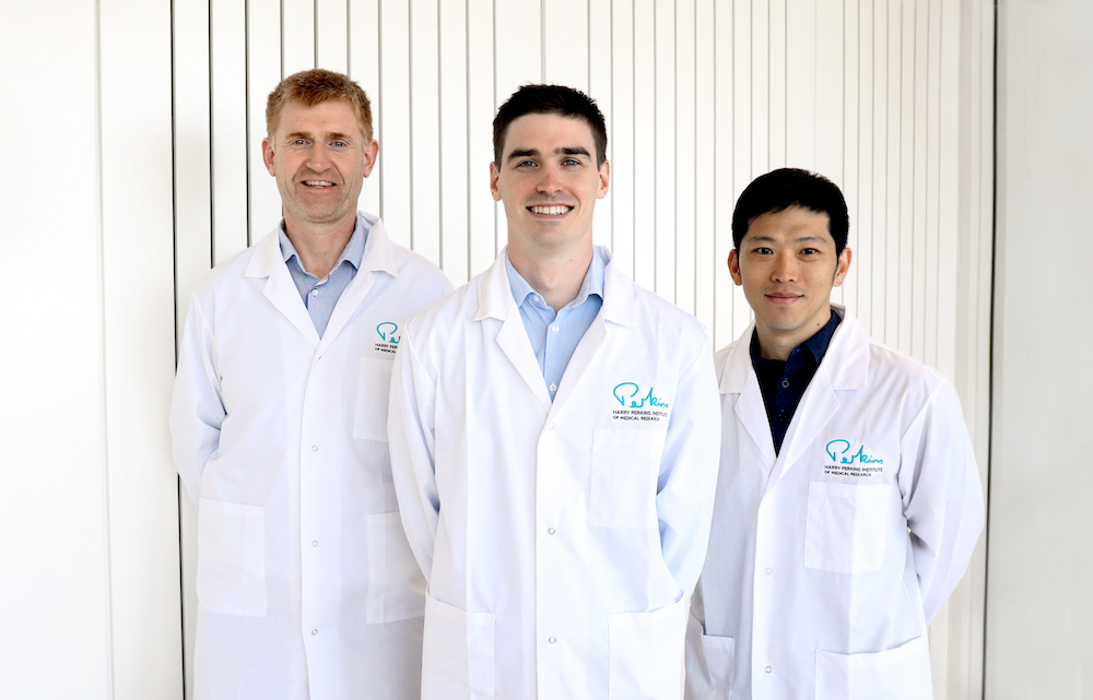
Matt Hepburn, from the Perkins and UWA’s BRITElab, said the team was trying to develop a tool that could measure cell stiffness, which has been implicated in many diseases including cardiovascular disease, muscular dystrophies, and the onset and progression of cancer.
“Cells pull on their environment to move around, grow and reproduce. When the cell pulls on its surroundings the stiffness of the cell’s environment acts as an important feedback loop to tell the cell where it is in the body and what to do next,” Mr Hepburn said.
BRITElab head, Dr Brendan Kennedy, said his team had developed a quantitative imaging model that was capable of displaying the stiffness of a cell’s environment in 3D for the first time – opening the door to innovative ways to study cells during disease progression.
“Deeper understanding into the connection between stiffness and cell behaviour has the potential to lead to new strategies to treat disease from an entirely new angle,” Dr Kennedy said.
“Our development could help biologists study the effect of stiffness on cell behaviour, and better understand how the knowledge gained from studying 2D models translates to cells in a 3D environment that more closely resembles the conditions found in our bodies.”
“Our developments could lead to new cures and treatments for diseases such as muscular dystrophies and cancer by helping uncover new behaviours in cells that could be targeted with drugs,” Dr Kennedy said.
The study looked at both healthy cells as well as cells that displayed an increase in some of the disease signals seen in breast cancer.
When studied in a 3D environment, the research showed that compared to healthy cells, cancerous cells exhibited higher levels of stiffness in their surroundings and the tool was able to differentiate between the different cell types.
UWA’s Dr Yu Suk Choi said that the technology could help answer many questions about cell behaviour and disease that were previously unanswerable.
“We believe that our research demonstrates that we can now use this quantitative model to study the impact of stiffness on cell biology in conditions that better mimic those found in the body,” Dr Choi said.
“Now we can study cells in a range of conditions to better understand the influence of stiffness on how different diseases start and progress.”
Perkins Director, Professor Peter Leedman, said this study was a great demonstration of the breakthrough research happening in Western Australia.
“Our collaborative teams of researchers are making discoveries here in Perth that could improve the way scientists around the world investigate health and disease,” Professor Leedman said.
The researchers said they next planned to collaborate with teams of biologists, medical researchers and engineers to investigate more, differing cell types and study changes in cell behaviour when the stiffness of the cell environment was closely controlled.
The study was published in Biomedical Optics Express, a top peer-reviewed journal that covers areas of biomedical optics, radiology and medical imaging.
